
Full blood count (FBC)
The full blood count (FBC) is a commonly ordered test and is is often considered a routine request for all in-patient admissions. Among outpatients it is likely the most commonly ordered test. The FBC includes information on;
- Haemoglobin (HB)
- Red cell parameters (RBC, MCV, MCH, MCHC, RDW)
- Total white blood cell count (TWBC)
- Platelet count (PLT)

Additional examinations may be be performed on top of the FBC. These include white cell differential count (DC) and reticulocyte count (RETIC). A morphological examination of the blood smear, also known as peripheral blood film (PBF) examination may also be performed if there are abnormalities identified on the FBC.
Haemoglobin (HGB)
The concentration of haemoglobin in blood is measured spectrophotometrically. Red cells are lysed using chemical detergents which breakdown the red cell membrane and release the haemoglobin contents into solution. A photometer measures the absorbance of the haemoglobin solution at a specific wavelength which directly correlates with its concentration.
Normal haemoglobin concentration varies according to age and sex. Haemoglobin is highest in the newborn and then gradually declines during the first 6 months of life and thereafter remains low throughout childhood. Levels increment at puberty with the spurt higher in males as compared to females. Adult males continue to have higher haemoglobin than females until the later stages of life when the haemoglobin level is somewhat equal between male and female. A person is considered to have anaemia when their haemoglobin is below the reference range for their age and gender.

http://www.who.int/vmnis/indicators/haemoglobin.pdf
Counting red cells
Modern automated haematology analysers are capable of counting and sizing red cells accurately. Measuring the number of red cells in a fixed volume and measuring the size of each cell is usually done using an impedance measurement method. This method is also known as the Coulter principle, named after the person who described this measurement technique.
Haemocytometer
In the ‘olden days’, counting of red cells was performed using a haemocytometer. This technique is laborious, inefficient and its accuracy is hight operator dependant as can be seen in the two videos below.
Impedance counting
Impedance counting on the other hand can be fully automated. Modern high throughput automated instruments can perform counts on hundreds of samples within a couple of hours. The basic principles of impedance measurement of red cell counts and parameters are as explained in the videos below.
Red cell parameters
Most automated instruments are at least able to make two measurements on each red cell sampled, as explained in the previous video; red cell count and red cell size. Based on this measurement and plotting of the distribution histogram, various red cell parameters can be calculated which is useful when interpreting results and making a clinical diagnosis.
Red blood count (RBC) is a measure of the number of red cells in a unit volume of blood. Normal RBC varies according to age and sex. The normal range is usually between 4.7 – 6.1 x 10¹²/L in males and 4.2 – 5.4 x 10¹²/L in females.
RBC is reduced in most cases of anaemia associated with diminished erythropoiesis (e.g. aplastic anaemia, iron deficiency) or increased destruction (e.g. haemolytic anaemia). However, in carriers of thalassaemia and certain haemoglobinopathies, the RBC can be inappropriately high despite the anaemia. RBC therefore is a useful parameter to distinguish microcytic anaemia due to iron deficiency from thalassaemia.
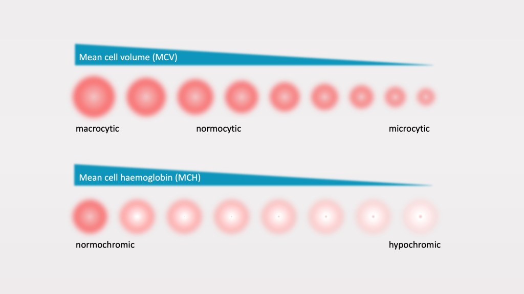
Mean cell volume (MCV) is a measure of the average volume of red cells in a blood sample. Low MCV denotes microcytosis (small red cells) while a high MCV denotes macrocytosis (large red cells). MCV therefore is often used to classify anaemias according to whether it is a microcytic, normocytic or macrocytic anaemia. You will learn more on the classification of anaemia in subsequent modules.
MCV varies according to age. In adults the MCV is normally 80 – 100 fL. Newborns have higher MCV while children have lower MCV as compared to adults. There is no significant MCV difference between males and females.
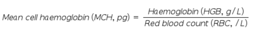
Mean cell haemoglobin (MCH) is a calculated parameter. It reflects the average amount of haemoglobin present in red cells of a patient sample. A low MCH denotes hypochromia and in anaemia is usually associated with low MCV (hypochromic microcytic anaemia). Normal MCH range in the adult is 27 – 33 pg.
Mean cell haemoglobin (MCH) is a calculated parameter. It reflects the average amount of haemoglobin present in red cells of a patient sample. A low MCH denotes hypochromia and in anaemia is usually associated with low MCV (hypochromic microcytic anaemia). Normal MCH range in the adult is 27 – 33 pg.
Mean cell haemoglobin concentration (MCHC) is the estimated mean concentration of haemoglobin in the red cells of a blood sample. MCHC is not a particularly useful independent red cell parameter for diagnostic interpretation. It may however be elevated when there is red cell spherocytosis (where red cells are spherical instead of being discoid) and therefore can be a useful clue when this condition is suspected.

Reticulocyte
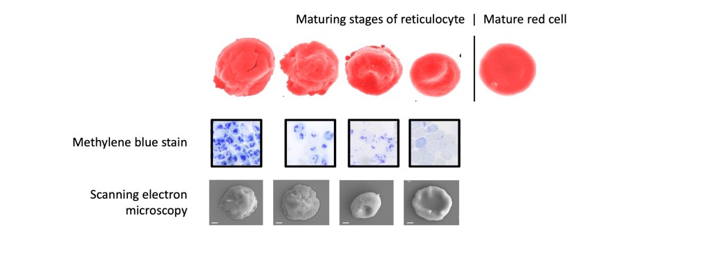
Reticulocytes are immature non-nucleated red cells that are present in circulation. In the normal subject, 1-2% of circulating red cells are reticulocytes. Reticulocytes are distinguished from other red cells by the presence of remnant RNA which can be identified by supravital stain or nucleic acid binding fluorescent dyes.
Reticulocytes are especially useful as a marker of marrow activity. Production failure in the marrow (e.g. aplastic anaemia, dyserythropoietic anaemia) causes reduced reticulocyte count while hyperactivity or stimulation of the marrow due to conditions such as bleeding and haemolysis causes an increased reticulocyte count.
Red cell morphology
In addition to quantitative measurements as described above, red cells can also be assessed based on its appearance. This is usually performed by preparing a blood smear, staining, and examining the cells under a microscope. This procedure is called a peripheral blood film (PBF) examination.
Variations of cell sizes and shapes as visualised under the microscope may give clues to the patient’s underlying condition or diagnosis. Some common red cell morphological abnormalities and their associated conditions are shown below.

The following video meanwhile illustrates how the peripheral blood film may provide important clues to the diagnosis of various anaemias
